KUROKI Medical Software
โปรแกรมวิเคราะห์และประมวลผล สำหรับชีววิทยาและงานทางการแพทย์ ยี่ห้อคุโรกิ
BIOWIZARD Ver. 4.1 is a new generation image analysis software meant for scientist to do analysis in the simplest way. This is a single screen window based system. The system is flexible, independent to adopt any capture card, camera and Microscopes.
BIOWIZARD can handle monochrome (8 bits) and color (24 bits) images. Multiple images of any size can be opened and displayed on the screen for analysis or comparison. The software support most common formats BMP, JPEG, TIFF, PNG, and GIP & PSD. The full screen real time image can be observed and captured on the same platform. Since system is made in window environment, graphs and charts displayed on the monitor can be quickly transferred into other window-based program like MS Word, MS Excel or any other commercial window based software for the use in reports and presentation.
MEASUREMENTS
- Sportial Calibration
- Line Measurements for Distance, Length, Width, Perimeter, Angle, Three point Radius.
- Area by enclosed line controlled by four arrow keys available on the keyboard arrow with zoomed preview. The Line measurement is not effected on zoomed images.
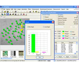
COUNT & CLASSIFICATION
Identification of objects in an image, count them, obtain several features measurements. Objects identification by user or automatically. User defined classification on basis of size or intensity.
THRESHOLD PARTICLE
Manual, Auto bright and Auto dark methods to identify intensity range defined object to be measured. Various calculation & measurements available for selected Particle are; Dimensions, Area, Perimeter, Ferrite Length, Min/ Max Radius, Thread Length, Thread Width, Fiber Length, Fiber Width.
MORPHOMETERY
Roundness, shape, Orientation, Elongation, Equal Circular Diameter, Equal Sphere Volume.
LOCATIONAL
Centroid X, Centroid Y, Major X1, MajorY1, Minor X1, MinorY1, Major X2, MajorY, Major X2, MinorY2, Box X1, X2, Box Y, Y2 & Box Area. Measure area fraction & Volume fraction. Identify multiple phases within Microstructure. Also delineate phases from the histogram.
- Three Options: Direct Print out with Original image & Tabular Results.
- Export to MS Office
- Excel for further modification.
For more information please contact : info@kurokicorp.com